Osteoarthritis (OA) is also known as degenerative joint disease. It is the most common type of arthritis. It occurs when the cartilage at the joint surface breaks down and the underlying bone becomes damaged.
Symptoms include pain and stiffness. Crepitus can be present; this is a grating or grinding sensation as the joint moves through its normal range. Swelling may also occur. Symptoms vary from person to person and some people report their symptoms to worsen with damp weather and/or activity. These symptoms result in restricted mobility and can lead to the muscles surrounding the joint becoming weaker, reducing the stability of the joint.
OA can be diagnosed by a physical examination. MRI scans or x-rays can also be used to determine the specific degeneration at a joint.
Treatment for OA varies depending on the severity of the joint degeneration and the pain this is causing. Mild OA can be managed with painkillers, physiotherapy, and steroid injections.
Arthroscopic surgery may be indicated to wash out a joint and remove loose fragments of bone that cause a joint to lock.
If a joint has severely degenerated, joint replacement surgery will be indicated. In the UK, hip and knee replacements are the most common types of joint replacement surgery.
Orthotic treatment may be used at any stage of osteoarthritic disease.
Foot orthotics are used to provide stability, support and to slow the progression of the disease at the bones of the mid and forefoot or ankle. They can also be used to influence the ground reaction forces acting at the knee and hip. This can be an effective treatment in the early stages of OA at these joints.
In more severe cases of OA in the foot and ankle, orthotic footwear and insoles can be used to offload painful joints and accommodate severe joint deformity.
Hinged SMOs (supra malleolar orthoses) are lightweight, neat plastic ankle braces. They can be used in severe OA at the ankle where there has been the collapse of the joints. These provide stability and relieve pain to allow for continued mobility.
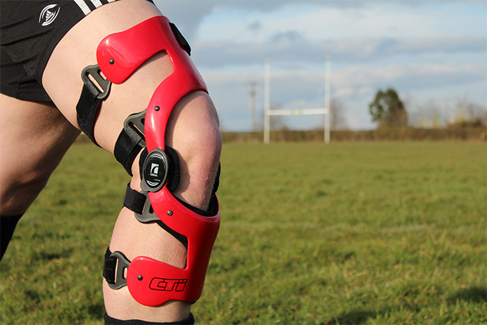
Knee braces can be a very effective way of treating unilateral compartmental OA at the knee. This type of bracing offloads the compartment of the knee that is degenerating, reducing pain and progression of the disease. It is also an alternative treatment in severe OA when surgery is not indicated. Research and development has moved forward in recent years with knee bracing for this condition and it is now recognised as a very effective form of treatment.
Each case of OA presents itself very differently in terms of the symptoms and the biomechanical deficits the joint disease is causing. At LOC, we provide a detailed orthotic assessment and have vast experience in prescribing effective orthotic treatment for this painful condition.
We now have the advantage of using our Gait Lab facilities to test and provide objective measurements of the effectiveness of joint offloading orthotics.
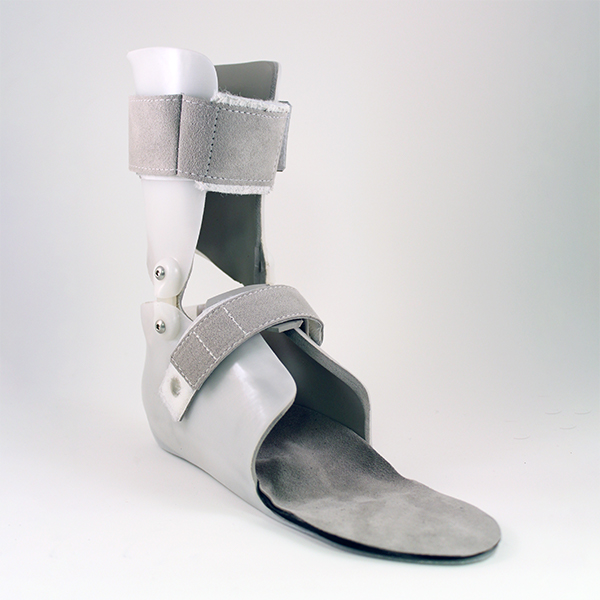
Above: SMO
An AFO is an Ankle Foot Orthosis which as the name would suggest encompasses the ankle and foot. The objective is to control the position and movement of the ankle. AFOs are used to support weak limbs; they can also be used to immobilise the ankle and lower leg to correct foot drop. When set up correctly they can also have a great influence on the knee and hip joints. They are the most commonly used Orthoses.
The length of time that one needs to wear an AFO very much depends on the condition being treated. If it is a long-term condition like cerebral palsy or post-polio syndrome it is likely to be years as the condition cannot be cured. Your orthotist will advise you.
A patient’s comfort in their AFO is vital for compliance with the prescribed wearing regime.
So there are a number of steps the orthotist should take to ensure a comfortable fit: the patient’s heel should fit fully into the heel cup without excess space, the contours of the plantar surface of the AFO should match the patient’s foot, for children there needs to be up to half an inch growth room in the toe shelf length. At LOC we use our Gait Laboratories at our Kingston and Manchester clinics to fine-tune our bespoke orthotics.
A GRAFO is used to control instabilities in the lower limb by maintaining proper alignment of limbs and controlling their motion. It reaches around to the front of the knee extending down to the ankle. It works by altering a patient’s limb presentation to displace load and impact as well as offering further control to the knee.
The cost of an AFO is dependent on the type of AFO that has been prescribed and the material that it has been made with. Carbon fibre will be more expensive than metal or plastic for example. LOC’s bespoke AFOs cost can be found on our Orthotic Prices page.
The ability to drive while wearing an AFO is dependent on the condition being treated and the orthosis that has been prescribed. If wearing a hinged AFO, for example, you will be able to drive, but if wearing a knee brace, you won’t. Your orthotist will advise you.
The most flexible type of AFO is a Dynamic Ankle Foot Orthosis (DAFO). It is thin and provides flexible support to the foot and ankle.
Both normal AFOs and DAFOs improve static balance (eg: while standing). Research among MS sufferers suggests that DAFOs aided balance while walking more than AFOs.
The simple answer is: yes they can. However one has to be sensible and look for wide-fitting trousers/jeans preferably of light and thin material.
Typically an AFO is stiff and rigid whereas a DAFO is thin, flexible and wraps around the patient’s entire foot. A DAFO provides support but also allows some range of normal movement.
A Supra Malleolar Orthosis SMO gets its name from the part of the body it encompasses. Thus an SMO supports the leg just above the ankle bone or malleoli. It allows dorsiflexion and plantar flexion(toes up and toes down) but eliminates mediolateral movement.
It typically takes a few weeks but is slightly dependent on the chosen materials and current availability.

Sofia’s AFOs Help Her to Stand and Walk During Lockdown

LOC’s gait lab has helped Lilac to move independently

Sam Walmsley to explain OSKAR Framework to Neuro Conference 2021 Delegates

Meet John Turner – our Manchester Orthotics Lead

LOC to launch OSKAR Framework to CMSUK 2020 Delegates

OSKAR Clinic AFOs and gait laboratory are game-changers for cerebral palsy patient Austin

Cerebral palsy patient, Sophie, maintains independence during lockdown with LOC’s help